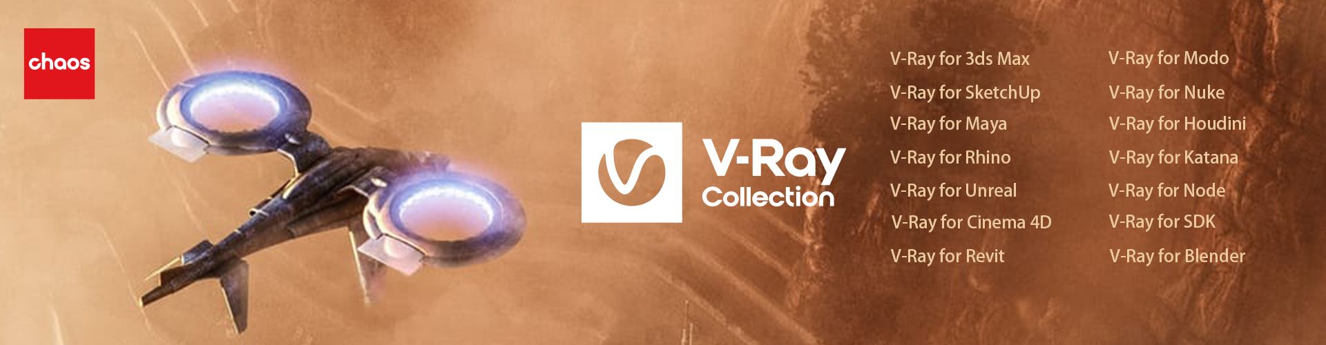
VRay是广受业界欢迎的渲染引擎
NUKE 是由The Foundry公司研发的一数码节点式合成软件
数字化设计、工程与娱乐软件服务和解决方案
Microsoft Windows操作系统和Microsoft Office系列软件
PTC解决方案来了! 2018-05-18
微软M365来了 2018-05-18
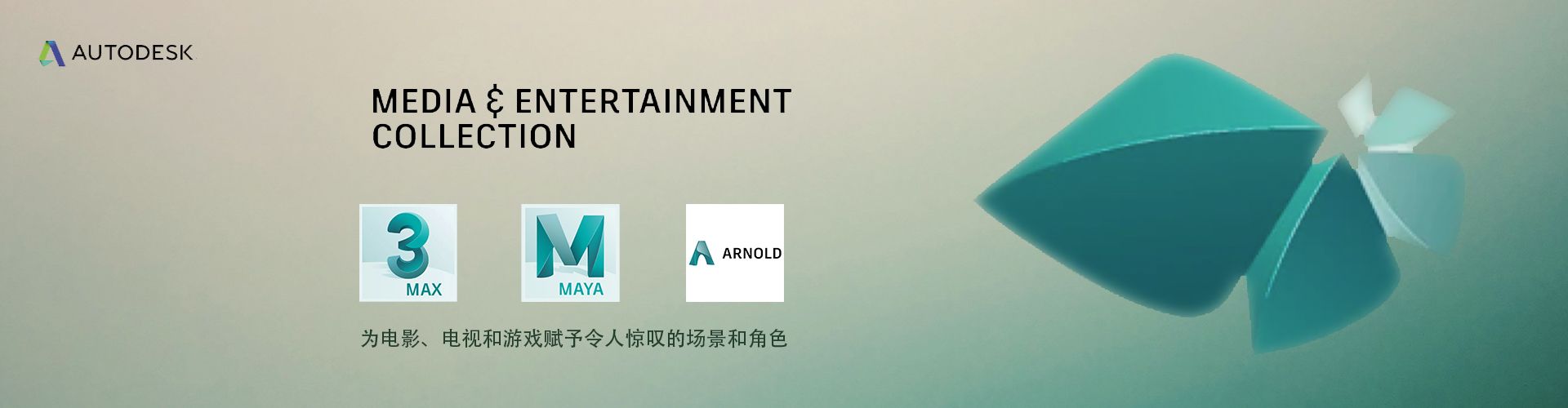
Autodesk 2022正式发布 2018-05-18
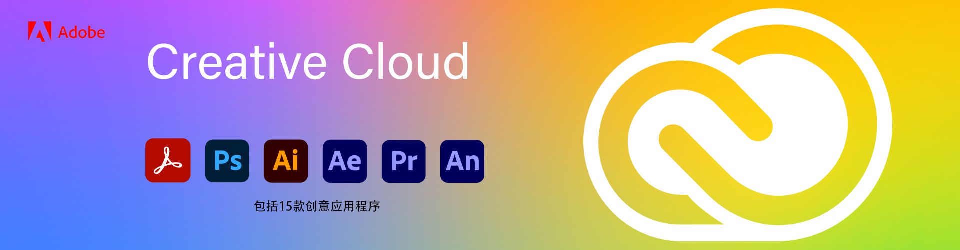
Adobe Creative Cloud 2022来了 2018-05-18